Home

Tulić się Zdarzenie Potomkowie ge 3d tomography Klasa dobrobyt Wyspy Owcze

Direct volume rendering: 3D visualization of the GE CT data with Amira... |  Download Scientific Diagram
Direct volume rendering: 3D visualization of the GE CT data with Amira... | Download Scientific Diagram

GE and NVIDIA Join Forces to Accelerate Artificial Intelligence Adoption in  Healthcare | NVIDIA Newsroom
GE and NVIDIA Join Forces to Accelerate Artificial Intelligence Adoption in Healthcare | NVIDIA Newsroom
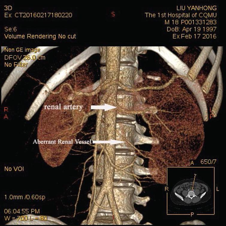
The Insight Zone: New Software Takes The 3D Printing Of Human Organs To A  New Dimension | GE News
The Insight Zone: New Software Takes The 3D Printing Of Human Organs To A New Dimension | GE News

CT scan - Wikipedia
CT scan - Wikipedia

Computed tomography angiography with 3D reconstruction in diagnosis of  hydronephrosis cause by aberrant renal vessel: A case report and mini  review - IOS Press
Computed tomography angiography with 3D reconstruction in diagnosis of hydronephrosis cause by aberrant renal vessel: A case report and mini review - IOS Press

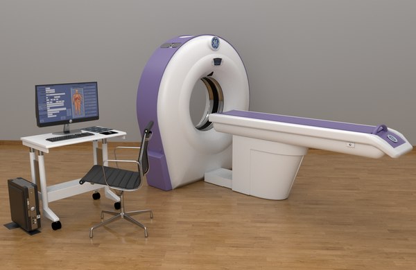
Ct machine brivo ct385 3D - TurboSquid 1345383

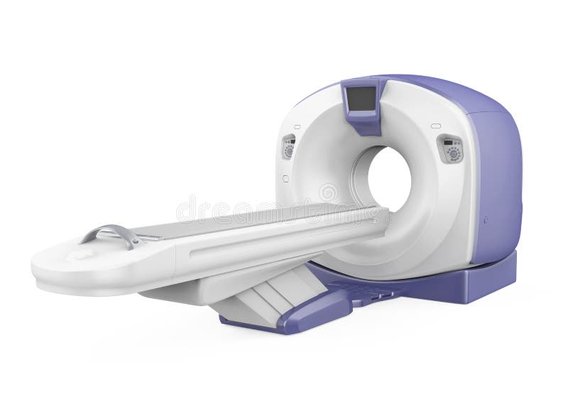
CT Scanner Tomography Isolated Stock Illustration - Illustration of render,  digital: 134859067
CT Scanner Tomography Isolated Stock Illustration - Illustration of render, digital: 134859067

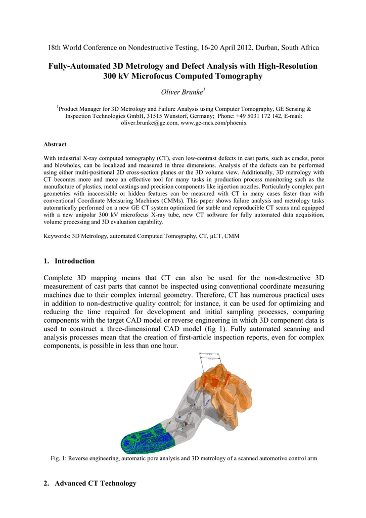
GE Inspection: Industrial Process Control with Computed Tomography (C…

X-ray computer Tomography (XRT / CT): Non-invasive 3D imaging of internal  structures - WUR
X-ray computer Tomography (XRT / CT): Non-invasive 3D imaging of internal structures - WUR

Tomographic ultrasound imaging (TUI) and 3D reconstructed intracranial... |  Download Scientific Diagram
Tomographic ultrasound imaging (TUI) and 3D reconstructed intracranial... | Download Scientific Diagram

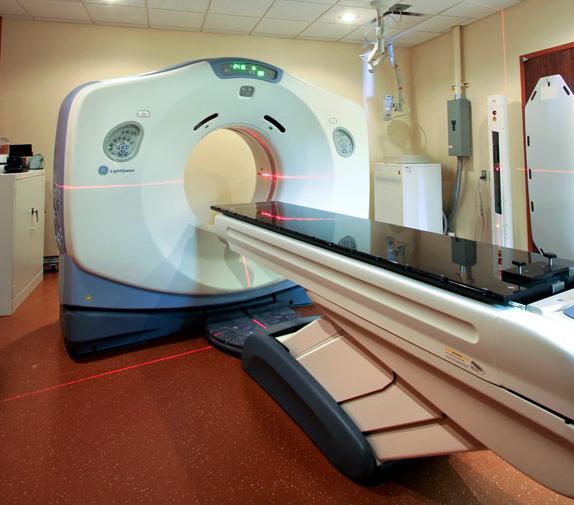
GE LightSpeed RT 16 CT Scanner - CT Medical Scanners

Phoenix Vtomex C: 3D CT Scanner | Industrial Metrology

3d Computed Tomography Angiography Of The Brain Stock Photo - Download  Image Now - iStock
3d Computed Tomography Angiography Of The Brain Stock Photo - Download Image Now - iStock

16- Slice New Ge Ct Scan Machine, Krystaal Healthcare India | ID:  22910021762
16- Slice New Ge Ct Scan Machine, Krystaal Healthcare India | ID: 22910021762

GE phoenix nanotom s Operating Manual | Manualzz

CT Scanner Tomography Isolated Stock Illustration - Illustration of  magnetic, computerized: 134859111
CT Scanner Tomography Isolated Stock Illustration - Illustration of magnetic, computerized: 134859111

Gadolinium-chelate-enhanced T1 GE 3D in the arterial phase shows a... |  Download Scientific Diagram
Gadolinium-chelate-enhanced T1 GE 3D in the arterial phase shows a... | Download Scientific Diagram

GE Healthcare Launches CT Scanner | DAIC

X-ray computed tomography system - Phoenix V|tome|x S240 - Waygate  Technologies - CT / high-resolution / 3D
X-ray computed tomography system - Phoenix V|tome|x S240 - Waygate Technologies - CT / high-resolution / 3D
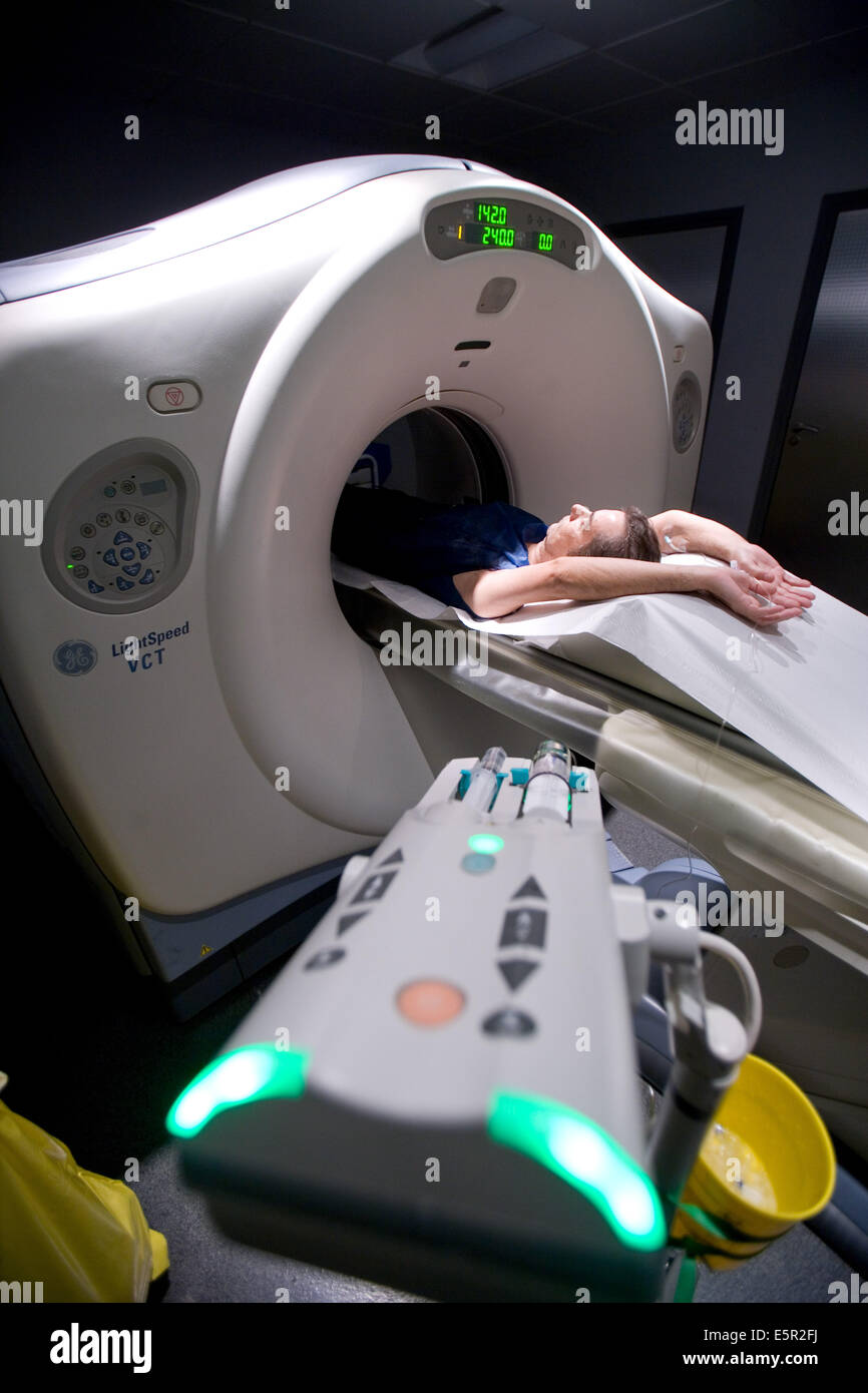
GE's super-fast CT scanner could save lives, definitely makes gory GIFs |  Engadget
GE's super-fast CT scanner could save lives, definitely makes gory GIFs | Engadget

Innova CT HD | GE Healthcare (United States)

Patient undergoing heart 3D CT scan, Department of Medical Imaging, Centre  Cardiologique du Nord, St Denis, France, At Stock Photo - Alamy
Patient undergoing heart 3D CT scan, Department of Medical Imaging, Centre Cardiologique du Nord, St Denis, France, At Stock Photo - Alamy
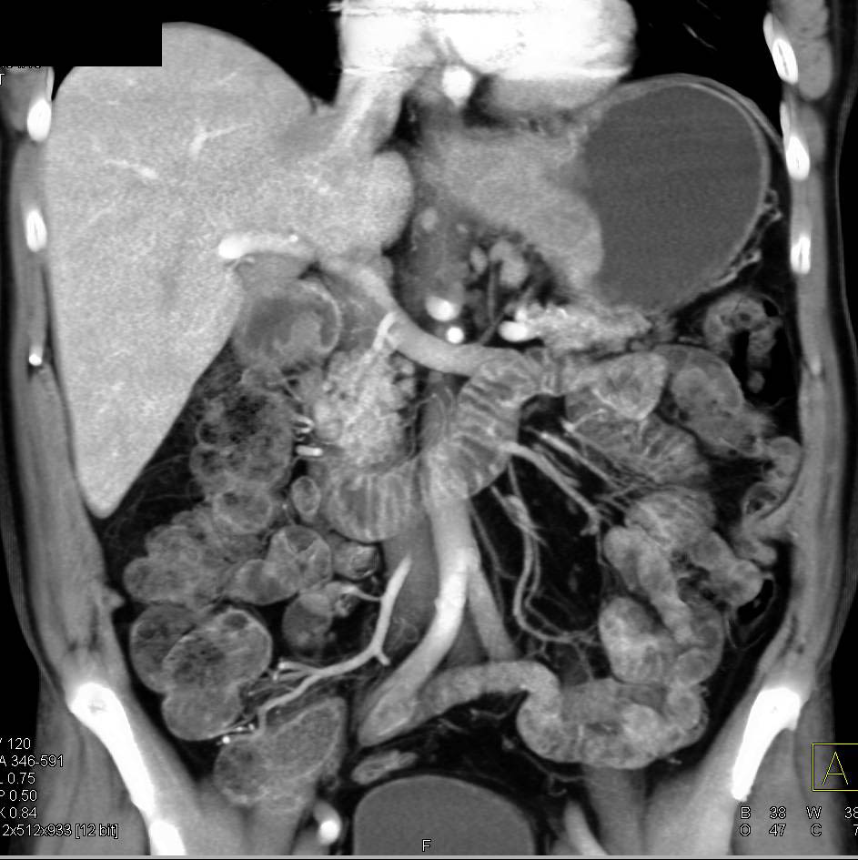
Impressive 3D Mapping of Adenocarcinoma of Gastroesophageal (GE) Junction  with Celiac Nodes. Tumor Extent Best Defined on 3D VRT Images - Stomach  Case Studies - CTisus CT Scanning
Impressive 3D Mapping of Adenocarcinoma of Gastroesophageal (GE) Junction with Celiac Nodes. Tumor Extent Best Defined on 3D VRT Images - Stomach Case Studies - CTisus CT Scanning