Home

Imperializm Obsługiwać drut light or heavy labeled amino acids klasa import Hodowla

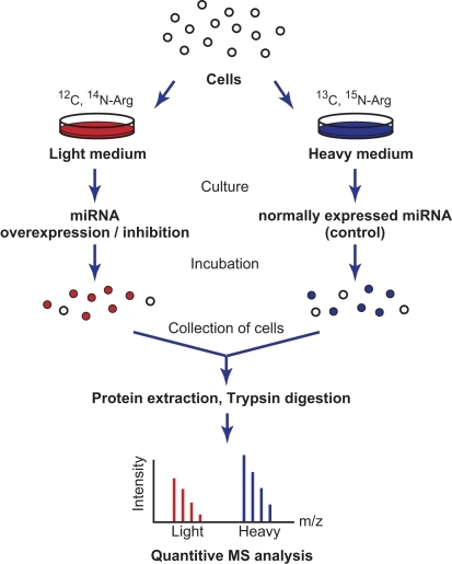
IJMS | Free Full-Text | SILAC-Based Quantification of TGFBR2-Regulated  Protein Expression in Extracellular Vesicles of Microsatellite Unstable  Colorectal Cancers | HTML
IJMS | Free Full-Text | SILAC-Based Quantification of TGFBR2-Regulated Protein Expression in Extracellular Vesicles of Microsatellite Unstable Colorectal Cancers | HTML

QPrEST Standards for Absolute Protein Quantification using Mass  Spectrometry - Atlas Antibodies
QPrEST Standards for Absolute Protein Quantification using Mass Spectrometry - Atlas Antibodies

Figure 14 from Stable-isotope labeling by amino acids in cell culture (  SILAC ) | Semantic Scholar
Figure 14 from Stable-isotope labeling by amino acids in cell culture ( SILAC ) | Semantic Scholar

Quantitative proteomics by amino acid labeling in C. elegans | Nature  Methods
Quantitative proteomics by amino acid labeling in C. elegans | Nature Methods

Stable Isotope Labeling with Amino Acids in Cell Culture (SILAC) for  Studying Dynamics of Protein Abundance and Posttranslational Modifications
Stable Isotope Labeling with Amino Acids in Cell Culture (SILAC) for Studying Dynamics of Protein Abundance and Posttranslational Modifications

Intercellular Transfer of Proteins as Identified by Stable Isotope Labeling  of Amino Acids in Cell Culture* - Journal of Biological Chemistry
Intercellular Transfer of Proteins as Identified by Stable Isotope Labeling of Amino Acids in Cell Culture* - Journal of Biological Chemistry
Schematic representation of SILAC labeling and proteome | Open-i

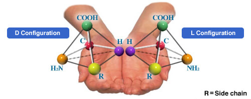
What are L-, D-, and DL-Amino Acids? | Enhancing Life with Amino Acids |  About us | Ajinomoto Group Global Website - Eat Well, Live Well.
What are L-, D-, and DL-Amino Acids? | Enhancing Life with Amino Acids | About us | Ajinomoto Group Global Website - Eat Well, Live Well.

Stable isotope labeling by amino acids in cell culture - Wikipedia

Figure 3 | Proteomics progresses in microbial physiology and clinical  antimicrobial therapy | SpringerLink
Figure 3 | Proteomics progresses in microbial physiology and clinical antimicrobial therapy | SpringerLink

SILAC-based Proteomics Analysis Service - Creative Proteomics

Light or heavy isotope labeled amino acids (Lys, +6 Da; Arg, +10 Da)... |  Download Scientific Diagram
Light or heavy isotope labeled amino acids (Lys, +6 Da; Arg, +10 Da)... | Download Scientific Diagram

Highlighted methods publication: Finding protein functions by quantitative  mass spectrometry
Highlighted methods publication: Finding protein functions by quantitative mass spectrometry

Characterization of the Tyrosine Kinase-Regulated Proteome in Breast Cancer  by Combined use of RNA interference (RNAi) and Stable Isotope Labeling with Amino  Acids in Cell Culture (SILAC) Quantitative Proteomics* - Molecular &
Characterization of the Tyrosine Kinase-Regulated Proteome in Breast Cancer by Combined use of RNA interference (RNAi) and Stable Isotope Labeling with Amino Acids in Cell Culture (SILAC) Quantitative Proteomics* - Molecular &

Stable Isotopic Labeling by Amino Acids in Cell Culture or SILAC <  Proteomics
Stable Isotopic Labeling by Amino Acids in Cell Culture or SILAC < Proteomics

Cells can be metabolically labeled with a combination of Aha and/or... |  Download Scientific Diagram
Cells can be metabolically labeled with a combination of Aha and/or... | Download Scientific Diagram

1) Natural amino acids, (2) N-terminus heavy and light chain, (3)... |  Download Scientific Diagram
1) Natural amino acids, (2) N-terminus heavy and light chain, (3)... | Download Scientific Diagram

Amino acid based labeling techniques. (A) SILAC labeling. Proteins are... |  Download Scientific Diagram
Amino acid based labeling techniques. (A) SILAC labeling. Proteins are... | Download Scientific Diagram

Quantitative Mass Spectrometry-based Proteomics | IntechOpen

Comparison of Isotope-labeled Amino Acid Incorporation Rates (CILAIR)  Provides a Quantitative Method to Study Tissue Secretomes - Molecular &  Cellular Proteomics
Comparison of Isotope-labeled Amino Acid Incorporation Rates (CILAIR) Provides a Quantitative Method to Study Tissue Secretomes - Molecular & Cellular Proteomics

Stable Isotope Labeling by Amino Acids in Cell Culture for Quantitative  Proteomics | SpringerLink
Stable Isotope Labeling by Amino Acids in Cell Culture for Quantitative Proteomics | SpringerLink

Encoding quantitative information into whole proteomes with SILAC.Cells...  | Download Scientific Diagram
Encoding quantitative information into whole proteomes with SILAC.Cells... | Download Scientific Diagram

Quantitative, Time-Resolved Proteomic Analysis by Combining Bioorthogonal  Noncanonical Amino Acid Tagging and Pulsed Stable Isotope Labeling by Amino  Acids in Cell Culture* - Molecular & Cellular Proteomics
Quantitative, Time-Resolved Proteomic Analysis by Combining Bioorthogonal Noncanonical Amino Acid Tagging and Pulsed Stable Isotope Labeling by Amino Acids in Cell Culture* - Molecular & Cellular Proteomics

Light or heavy isotope labeled amino acids (Lys, +6 Da; Arg, +10 Da)... |  Download Scientific Diagram
Light or heavy isotope labeled amino acids (Lys, +6 Da; Arg, +10 Da)... | Download Scientific Diagram

Schematic representation of a stable isotope labelling with amino acids...  | Download Scientific Diagram
Schematic representation of a stable isotope labelling with amino acids... | Download Scientific Diagram

Light or heavy isotope labeled amino acids (Lys, +6 Da; Arg, +10 Da)... |  Download Scientific Diagram
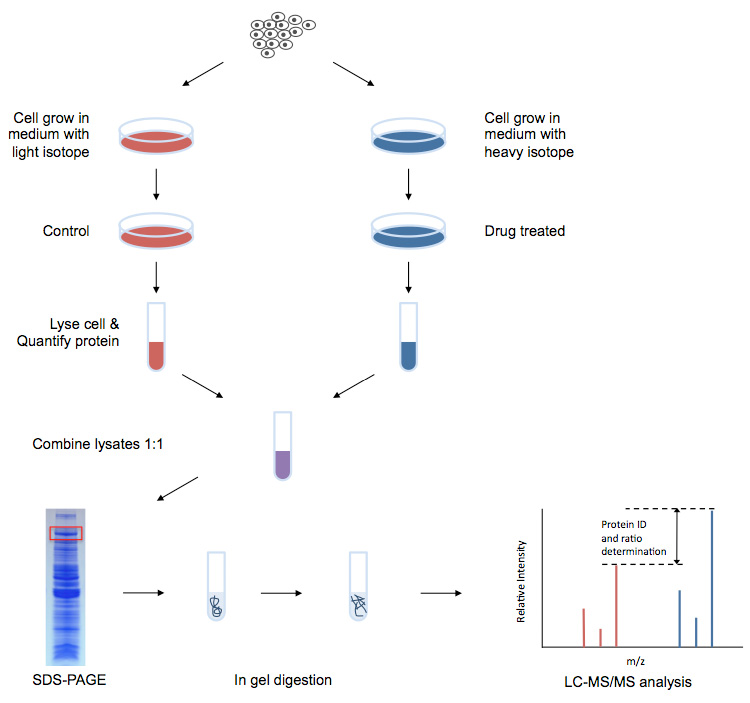
Light or heavy isotope labeled amino acids (Lys, +6 Da; Arg, +10 Da)... | Download Scientific Diagram